|
|
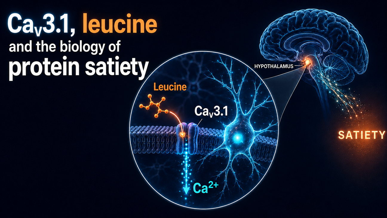
Researchers have uncovered how the brain may translate a protein-rich meal into a feeling of fullness. A study published in Cell Metabolism identifies CaV3.1—a calcium channel in the hypothalamus—as a direct sensor of leucine, an amino acid abundant in dietary protein
Leucine binds to a specific pocket in CaV3.1 and lowers the voltage threshold required to activate the channel. This initiates calcium influx, which is amplified by TRPC5 and depolarizes appetite-suppressing pro-opiomelanocortin (POMC) neurons. The resulting pathway provides a molecular explanation for how dietary protein can reduce food intake.
Alomone Labs products helped researchers characterize key stages of this mechanism. The selective in CaV3.1 inhibitor (Rac)-TTA-P2 (#T-155) abolished leucine-induced activation in mouse primary POMC neurons, human iPSC-derived POMC neurons, and mouse brain slices, demonstrating that in CaV3.1 activity is required for the response. The study also used the Anti-CACNA1G (in CaV3.1 ) Antibody (#ACC-021) and Anti-TRPC5 Antibody (#ACC-020) to investigate the channels involved in the pathway. In complementary immunoprecipitation-based binding experiments, the Anti-CACNA1G antibody helped demonstrate that leucine binds directly and specifically to in CaV3.1 .
In mice, deleting CaV3.1 from the relevant hypothalamic neurons abolished leucine-induced appetite suppression and disrupted the satiety normally associated with a high-protein diet. Conversely, activating the channel reduced food intake and body weight while enhancing the effects of the GLP-1 receptor agonist liraglutide.
While the study used both mouse- and human-derived models to establish in CaV3.1 ’s role in leucine-induced POMC neuron activation, its feeding and metabolic effects were evaluated only in male mice. Further research is therefore needed to explore potential sex-dependent differences and determine whether the mechanism contributes to appetite regulation in humans.
Because CaV3.1 is widely expressed throughout the brain, leucine sensing in other CaV3.1 -expressing cells may also have physiological functions beyond the control of food intake.
👉 Read the blog here.
|
|
|
|
CaV3.1 Channel Identified as the Brain’s Protein Satiety Sensor
|
|
Learn how a calcium channel helps the brain turn dietary protein into a satiety signal.
|
|
|
|
|
|
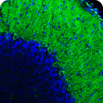
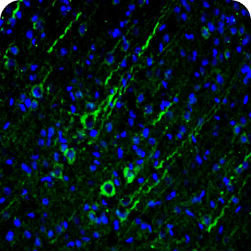
A highly cited, knockout-validated antibody for detecting the T-type calcium channel CaV3.1 in human, mouse, and rat samples. It is a valuable tool for studying CaV 3.1 expression in neuronal excitability, rhythmic firing, sleep regulation, pain, epilepsy, cardiac function, and neuroendocrine and metabolic processes, including appetite control. Validated for Western blot and immunohistochemistry.
|
|
|
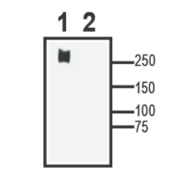
A membrane protein extract from Xenopus oocytes overexpressing rat CACNA1G (CaV 3.1). Designed as a target-specific positive control for Western blot, it helps confirm antibody performance, band size, and assay conditions.
|
|
|
A highly cited, knockout-validated antibody for detecting the calcium-permeable cation channel TRPC5 in human, mouse, and rat samples. It is a valuable tool for studying TRPC5 expression in neuronal excitability, synaptic signaling, fear and anxiety, pain, appetite and metabolic regulation, and kidney function. Validated for Western blot and immunohistochemistry.
|
|
|
|
|

|
68 Citations
|
|
|
|
|
|
54 Citations
|
|
|
|
|
|
|
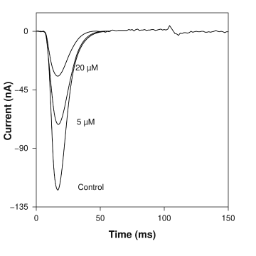
A highly pure, synthetic small molecule that functions as a potent and selective T-type voltage-gated calcium channel CaV 3.1 (CACNA1G) blocker. It is widely used as a pharmacological tool in neuroscience research to investigate neuronal excitability, synaptic transmission, and pathological conditions like epilepsy and chronic pain.
|
|
|
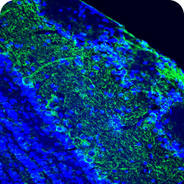
A highly specific antibody targeting an extracellular epitope of LEPR/OB-R, enabling cell-surface detection in live intact cells. It is suitable for research into leptin signaling, appetite and body-weight regulation, energy and glucose homeostasis, neuroendocrine function, and immune–metabolic interactions. The antibody recognizes mouse and rat and is validated for Western blot, IHC, ICC/IF, and flow cytometry.
|
|
|
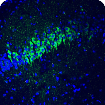
A highly specifc antibody for detecting 5-HT2C in human, mouse, and rat samples. It supports research into serotonergic signaling, appetite and body-weight regulation, mood, anxiety, reward, and other CNS functions. Validated for Western blot and immunohistochemistry.
|
|
|
|
|
|
63 Citations
|
|
|
|
|
1 Citation
|
|
|
|
|
|
|
We Continue to Fully Support Science Around the World
Leading membrane protein research tools for 35+ years.
We provide high-quality solutions for academia, pharma, biotech, and CROs – validated by over 30,000 scientific citations demonstrating our excellence.
|
|
|
|
|
|
|
|
|