Coronin-1A in Immune Cells
Coronin proteins, initially identified in Dictyostelium slime mold, are essential regulators of cellular motility and immune functions. In mammals, the coronin protein family includes five isoforms (coronins 1–5 or 1A, 1B, 2A, 2B, and 2C), all belonging to the WD40 repeat family, distinguished by a conserved seven-bladed β-propeller structure that facilitates critical protein-protein interactions. Notably, coronin 1a (also referred to as coronin 1) is restricted to cells of hematopoietic origin, such as lymphocytes, macrophages, and neutrophils. In the central nervous system (CNS), it is specifically expressed in microglia, the resident immune cells, which exhibit dynamic behaviors including surveillance, phagocytosis of pathogens and cellular debris, and interaction with surrounding cells. Microglial proliferation is markedly increased during CNS injury or pathology, making coronin-1A a reliable marker for their identification.
|
|
|
Biosensis’ Coronin-1A, Rabbit Polyclonal Antibody (R-1335-50) detects coronin-1A with high specificity by targeting the C-terminal peptide of human coronin-1A, chemically conjugated to keyhole limpet hemocyanin (KLH). It performs effectively in western blotting and tissue studies across various mammalian species, making it particularly useful for identifying microglia and investigating immune responses in the CNS.
Applications and Key Features
<![if !supportLists]>· <![endif]>Western Blotting: Sensitive detection of Coronin-1A in cell and tissue extracts.
<![if !supportLists]>· <![endif]>Immunohistochemistry: Accurate identification of microglia in tissue samples.
<![if !supportLists]>· <![endif]>Research Areas: Ideal for studying CNS immune responses, inflammation, and cellular damage.
|
|
|
|
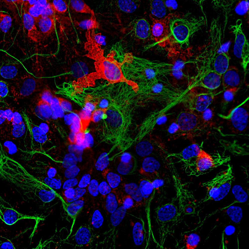
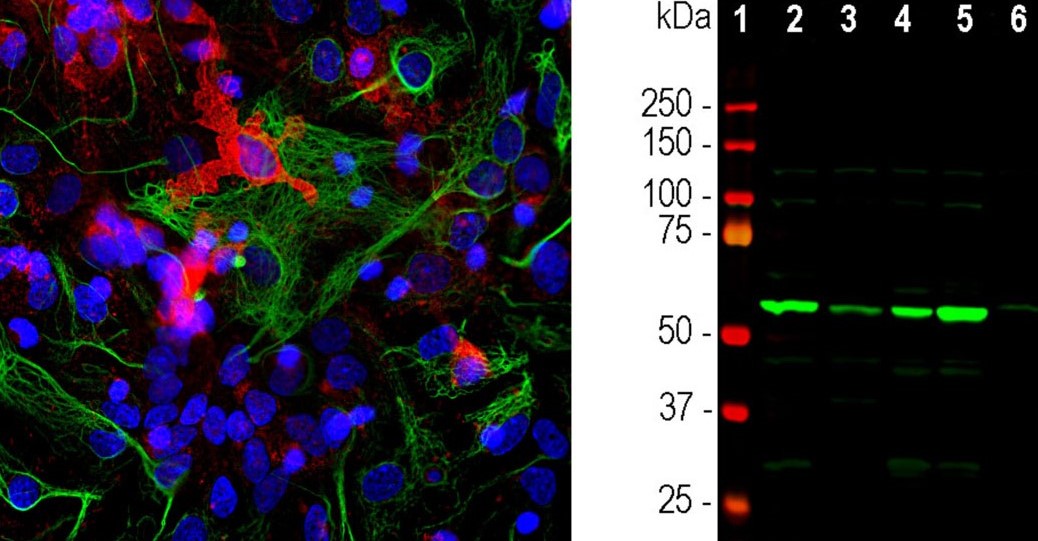
Left: Detection of Coronin 1a (R-1335-50, red) in cortical neuron-glial cell culture from E20 rat by Immunocytochemistry. Coronin 1a antibody was used at 1:1,000 dilution. Cells were co-stained with mouse antibody to GFAP (M-1375-100, green). Blue: Hoechst nuclear stain. The coronin-1A antibody labels protein expressed in the cytoplasm of microglia cells, while the GFAP antibody stains intermediate filaments in astrocytic cells.
Right: Western blot analysis of coronin-1A expression in tissue lysates using Rabbit polyclonal antibody to Coronin-1A (R-1335-50, green). [1] protein standard, [2] mouse brain, [3] rat brain, [4] cow cerebellum, [5] cow cortex, and [6] pig spinal cord. The strong single band about 55 kDa corresponds to the coronin 1a protein.
|
|

|
|
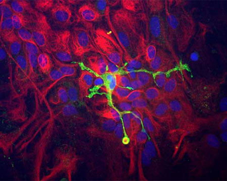
Immunocytochemistry of a mixed neuron/glial culture from newborn rat brain stained with Rabbit polyclonal antibody to Coronin-1A (R-1335-50, green) and Chicken polyclonal antibody to Vimentin (C-1409-50, red). Blue is nuclear DNA counter stain. Glial cells and fibroblasts stain with Vimentin, while microglia alone stain strongly and specifically for Coronin-1A, which can therefore be used as a robust marker of this important cell type.
|
|
|
|
Choose our R-1335-50 for Reliable and Accurate Research Results.
|
|
Biosensis is a company that is passionate about Science
|
|
|
|
|