|
|
| TrpV1: A Molecular Target in Pain and Inflammation Research
TrpV1 (Transient Receptor Potential Vanilloid 1) is a key molecular sensor for noxious stimuli and a pharmacological target for analgesic development. Its expression is upregulated in response to inflammatory mediators, making it a valuable marker in studies of neuropathic pain, inflammation, and thermoregulation. TrpV1 antibodies enable specific and sensitive detection of TrpV1 in both native and recombinant systems. |
|
|
Biosensis’ TrpV1 antibodies
Explore Thermal and Chemical Sensing
|
| Biosensis TrpV1 antibodies are a powerful research tool for studying pain perception, inflammation, and neurobiology. TrpV1 is a non-selective cation channel activated by capsaicin, heat, and protons, playing a key role in sensory signal transduction. Our antibodies offer consistent performance in detecting both native and denatured forms of TrpV1, making it suitable for a broad range of molecular and cellular biology applications.
They are validated for high specificity and sensitivity and are ideal for detecting endogenous TrpV1 expression across various sample types. |
|
| Key Features
· Host Species: Mouse
· Clonality: Monoclonal
· Species Reactivity: mouse and rat
· Predicted Reactivity: Guinea pig (based on sequence homology)
· Validation: Tested in WB, FC, IF, ICC and IHC
|
|
| Application Details:
· Western Blot: Detects a ~95–100 kDa band corresponding to TrpV1
· IHC/ICC: Suitable for fixed tissue or cultured cells; use with appropriate antigen retrieval methods
· Immunofluorescence (IF)
|
|
|
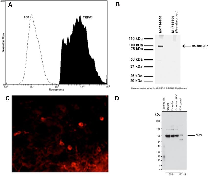
| A: Analysis of TrpV1 expression in rat PC12 cell line by Flow Cytometry.
B: Western blot of TrpV1 in rat PC12 cell lysates (80 µg/lane). M-1714-100 detects TrpV1 protein at 95-100 kDa.
C: Immunohistochemical staining of TrpV1 in mouse dorsal root ganglia. Immunoreactivity was visualized with anti-mouse-Cy3 conjugate (red).
Courtesy P. Vilimas, Flinders University Adelaide.
D: Western blot (denatured and reduced) of TrpV1 in cell lysates of forskolin and NGF stimulated 50B11 hybrid mouse x rat DRG cell lines and NGF-stimulated PC12 cells (10 µg/lane). M-1714-100 detects monomeric TrpV1 protein at 95-100 kDa.
Courtesy Dr. D. Matusica, Flinders University. |
|
|
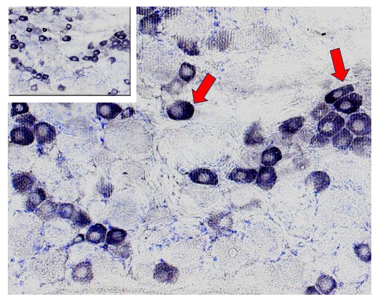
Analysis by IHC displaying the TrpV1-positive neurons (pseudounipolar ganglion cells, red arrows in enlargement) of dorsal root ganglia involved in pain sensation. Entire photo is presented in the inset image.
Immunohistochemistry generously provided by Dr. Szabolcs Takács & Dr. Erik Hrabovszky, Institute of Experimental Medicine, Budapest, Hungary. |
|
Key Features
· Host Species: Rabbit
· Clonality: Polyclonal
· Species Reactivity: Rat, Human
· Predicted Reactivity: Mouse (based on sequence homology)
· Validation: Demonstrated specificity via IHC analysis of rat dorsal root ganglia (DRG)
|
|
Application Details
· IHC (Frozen): 1:1000 – 1:2000 dilution
· Specifically validated for immunohistochemistry on frozen tissue sections
· Membrane-associated; expression enriched in sensory neurons
|
|
|
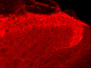
| Immunohistochemical staining of rat spinal cord using rabbit antibody to human capsaicin receptor. |
|
Key Features
· Host Species: Rabbit
· Clonality: Polyclonal
· Species Reactivity: Human, Rat
· Predicted Reactivity: Likely cross-reactive with other species due to sequence homology
· Validation: Specificity confirmed by IHC on frozen sections of rat dorsal root ganglia (DRG) and spinal cord
|
|
Application Details
· IHC (Frozen): 1:1000 – 1:2000 dilution
· This antibody has been validated in IHC-Frozen only.
· Predominantly membrane-associated, with possible cytoplasmic distribution
|
|
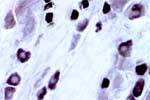
| Detection of capsaicin receptor in small neurons in rat dorsal root ganglion (DRG) using rabbit antibody to human capsaicin receptor (608-621): whole serum. |
|
Biosensis is a company that is passionate about Science
|
|
|
|
|